Why and when is treatment needed?
Reasons for treatment include
- • Symptoms associated with obstruction (pain)
• Reduced kidney function
• Development of stones or
• Infection
• High blood pressure (rarely).
What treatments are available?
The obstruction needs to be removed so that urine can pass freely from the kidney down to the bladder. This can be accomplished by several means:
• Cutting out the PUJ obstruction and joining the kidney onto the ureter (‘pyeloplasty’).
• Pyeloplasty i.e. cutting out the obstruction has the best results and lasts for the longest period. This can be achieved through a traditional surgery (‘open pyeloplasty’) or by keyhole surgery (‘robotic laparoscopic pyeloplasty’).
• Making a cut in the PUJ obstruction so that it splits open and becomes wider that way (‘endopyelotomy’).
• A cut in the PUJ obstruction (‘endopyelotomy’) is less effective than cutting it out altogether, but is possibly better than bursting it with a balloon. In some situations, it is dangerous because of neighbouring blood vessels.
• Bursting the obstruction with a balloon (‘balloon dilatation’)
Bursting the obstruction with a balloon is quick, the least invasive but is less effective and lasts for the shortest period. Furthermore, it produces scarring that can make corrective surgery more difficult. For some patients it is the best option because poor health makes other treatment dangerous.
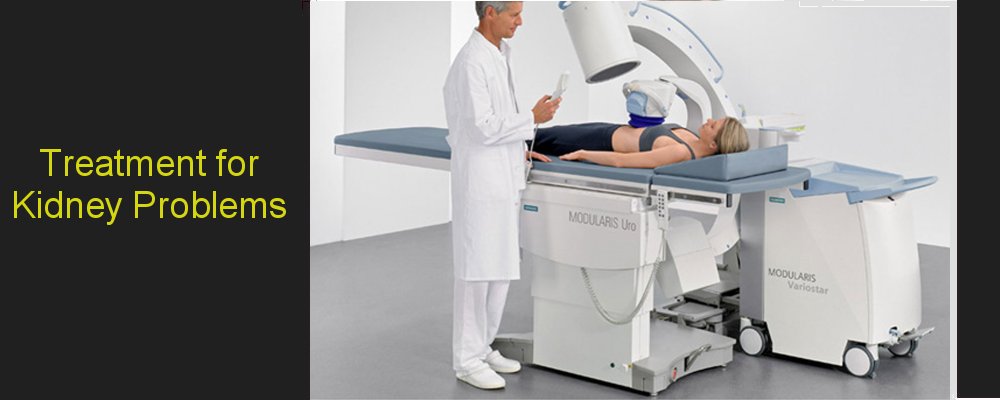
What is laparoscopic pyeloplasty surgery?
This is a key hole method for correcting the PUJ obstruction that avoids a large incision.
What is laparoscopy?
This is a technique to reach parts of the body without the use of large incisions. Instead, a narrow telescope and instruments are inserted through small incisions allowing surgery to be performed. The intention is to achieve the same results as would be obtained by conventional surgery.
What are the advantages of a pyeloplasty performed laparoscopically?
The advantages are multiple and include the following:
• smaller skin incision - four 1 cm incisions rather than a 30 cm incision
• better view because of the magnification of the system
• less pain because the incisions are smaller and the muscles are parted rather than cut
• 2 to 4 days in hospital compared to a week or longer by open surgery
• less blood loss and reduced need for a blood transfusion
• the ability to return to work in 2 to 4 weeks compared to 6 or more weeks after traditional open surgery
What are the disadvantages of a pyeloplasty performed laparoscopically?
As the hands are not directly in the body, it is less easy to feel what is happening compared to open surgery. In some situations, the tactile feedback can be important and if that becomes true, it would be necessary to make an incision to carry on. This is extremely rare. Other disadvantages include the increased length of time necessary for the operation, the significantly increased cost of the equipment and the necessity for the surgeon to be experienced in laparoscopy before being able to perform the operation.
How is a laparoscopic pyeloplasty performed?
After a general anaesthetic has been given, a telescope is placed through the urethra into the bladder. A little tube (stent) is placed in the ureter, which is the tube that connects the kidney to the bladder.
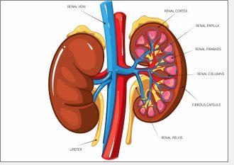
Afterwards, incisions are made in the side of the abdomen. Typically, there are about 3 or 4 incisions between 0.5 cm and 2 cm just below the ribs on the side of the problem. The narrow part of the junction between the renal pelvis and the ureter is excised. A new ‘join’ between the kidney and ureter is constructed. The operation lasts for about 2 hours to 3 hours. If there is a crossing vessel, the join is made on the other side of the crossing vessel and this makes the operation take a longer time to complete.
At the end of the procedure, there is usually a tube left inside the body near the site of the operation and this comes out through the skin (‘drain’). This is removed when fluid stops draining, which is usually after a day or so. There is another tube (‘catheter’) coming out from the bladder through the urethra and connected to a ‘catheter bag’. This is removed after a day or so also. The ‘stent’ placed internally between the kidney and the bladder remains at the end of the operation and is removed later under local anaesthetic about 6 weeks after surgery.
What are the side-effects of the laparoscopic pyeloplasty?
There are some risks associated with laparoscopy alone and some with the surgery. The common or serious risks of pyeloplasty include
• The operation does not work. This occurs in 5 to 10% of patients and would need a second procedure to correct. The risk of this is similar to that when performed by traditional open surgery
• A drain is required for a longer period than normal. In general, a drain is required for a day after the operation. If urine leaks for longer than expected, a drain may be necessary for a longer period
• Infection: this occurs rarely because of antibiotics, but the urine or wound can still become infected requiring further or different antibiotics
• Injury to other structures in the body. This is a risk of all surgery, but slightly higher when performed laparoscopically. Rarely, the kidney may have to be removed altogether
• Conversion from keyhole (laparoscopic) to traditional open surgery: if there is substantial difficulty performing the operation, then a traditional or larger incision may be required to complete the operation
• Bleeding may occur and a blood transfusion may have to be given. Rarely, the kidney may need to be removed or the bleeding controlled by special techniques
The risks of laparoscopy relate to the use of the small incisions and working with small instruments. These are rare include
• Entry into the abdomen instead of staying in retroperitoneum
• Gas entry into the skin around the incisions. This can result in the skin feeling crackly after surgery, but is short-lived
• Damage to nearby structures, which may include bowel or other organs in the abdomen. If this occurs, a large incision is necessary into the abdomen to fix this
Are there any problems urinating after treatment?
Immediately after treatment, there is usually a catheter in place. This is a tube that drains the bladder. After a day or so, the catheter may be removed if all is well and passing urine may be uncomfortable for a short period.
What can I expect post-operatively?
The drain and catheter are usually removed on the first or second day after surgery. You can usually go home between the second and fourth day of the operation. After 2 to 4 weeks after the operation, it may be possible to return to work. People vary and it depends on the degree of physical activity necessary to be performed and how you feel.
You can drive when you are able to brake safely, and this usually takes several weeks.
The internal tube (‘stent’) between the kidney and bladder is usually removed between three to six weeks after the operation. The stent can cause discomfort include pain on passing urine that may be felt in the back on the side of the operation, lower abdomen or tip of the penis. There may also be some blood in the urine. These problems can be worse if you are more active, but not always. The stent can usually be removed easily under a local anaesthetic by a special telescope inserted down the urethra i.e. the tube through which urine passes out of the bladder. This means coming into hospital for a morning or afternoon only and is performed as an outpatient procedure. Antibiotics will need to be taken for 3 days after the stent has been removed.
Three months later, another diuretic renogram (MAG3 study) is performed. This is another test performed as an outpatient, and will probably be similar to a test performed to help substantiate the diagnosis of PUJ obstruction in the first place. This is to determine whether obstruction is still present or not. Another study may be performed one year later.
JJ stents
A JJ stent is a specially designed hollow tube, made of a flexible plastic material that is placed in the ureter. The ureter is the natural tube that transmits urine from the kidney to the bladder. The length of the stents used in adult patients varies between 24 to 30 cm.
There are different types of stents, and some of these differences allow a stent to provide different benefits depending on the situation of the .
What's the reason for having a JJ stent?
A JJ stent may be placed for several reasons.
It allows urine to flow from the kidney to the bladder even when the ureter is blocked for one reason or another. This way, the kidney keeps working and is not damaged by being obstructed and avoids the severe pain that can occur when a kidney does not drain properly. The chance of an infection is also reduced significantly.
A stent protects the ureter and allows the ureter to heal even when damaged. If a stent is not placed and the ureter is hurt in some way or other, it can become too narrow when it heals forming what is called a stricture. Having a stent can prevent that from happening and makes it more likely that the ureter will work well afterwards.
Sometimes, a stent is placed because it makes a narrow ureter wider over a period of time. This can be important when access through the ureter is needed to pass instruments or remove stones. This would typically occur when an attempt to go up the ureter to get a stone has failed because it was too narrow. Inserting a stent makes it more likely that later attempts to get up the ureter will be successful.
What are the disadvantages of having a JJ stent?
It is not possible to predict who will or will not have side-effects with a stent. Some people tolerate stents without problems. Others find they have problems described below. Such problems may be present only at the beginning of having a stent and resolve over a few days or weeks. Other people may find their symptoms persist through out the period of the stent being present.
Stents can cause blood to appear in the urine at various times. Usually, physical activity of one kind or other results in movement of the stent inside the body. This can give rise to blood in the urine. Pain may be felt in the back (loin), bladder area, groin, penis in men or urethra in women, and sometimes the testicles. The discomfort or pain may be more noticeable after physical activities and after passing urine.
The stent can cause irritation of the bladder and so make it necessary to pass urine more frequently including the need to get up at night to pass urine. These symptoms can sometimes be improved by medication. Rarely, a stent may cause a woman to leak urine.
Once the stent has been removed, these side-effects go away
What problems can arise with a stent?
Sometimes, stents can become calcified and develop a coating similar to stones. Stents can also move out of position. When this happens, the stent usually moves into the bladder causing a deterioration in bladder symptoms i.e. going to the toilet to pass urine more frequently, discomfort in the area of the bladder and perhaps blood in the urine.
How does a stent interfere with daily life?
You can still go to work and play sports when you have a stent in place. However, you may feel more tired and experience discomfort during the day limiting your performance. In addition, you may need to visit a toilet more frequently and so need convenient access to a toilet.
Travel is possible, although medical attention may be required rarely. As stents can have side-effects, your ability to enjoy yourself may be limited as a result.
There are no restrictions on sexual activity, although there may be less enjoyment as a result of the side-effects described above.
What additional care is necessary when a stent is in place?
Drink at least 1½ to 2 litres (approximately four pints) of fluids a day
discuss with your doctor if you have troublesome side-effects
When might it be necessary to call a doctor?
You should contact a doctor if
• Constant and unbearable pain related to the stent
• Symptoms of a urine infection (fever, rigours, feeling unwell and pain passing urine
• The stent falls out
• There is a significant increase in the amount of blood in the urine
How is a stent inserted?
A stent is inserted usually under a general anaesthetic often in combination with another procedure depending on the reason for the stent. A telescope called a cystoscope is passed through the urethra (water pipe) and into the bladder. The stent is passed through the cystoscope and into the ureter. The position of the stent is checked with x-rays.
What alternatives are there to a JJ stent?
Sometimes, it may be reasonable not to leave a JJ stent if obstruction is likely to be transient. This may be risky and depends on the circumstances. If several procedures have been performed, there is often swelling making obstruction and pain a distinct possibility.
Occasionally, it may be possible to place a tube internally draining the kidney that comes out through the urethra (water pipe). This can simply be removed by pulling it out without needing a further procedure of any kind. The disadvantage is that it can remain for only a day or so.
Another alternative is to have tube placed directly through the skin and into the kidney. This is called a 'nephrostomy'. This is placed under guidance by ultrasound and the kidney has to be distended to get into the correct place without difficulty. As this is outside the body, it is slightly more inconvenient and can sometimes get pulled out by accident. Its advantage is that it usually drains better than a JJ stent which can be important if there is infection with obstruction of the kidney ('pyonephrosis').
How is a stent removed?
A JJ stent is removed with a cystoscope performed under local or general anaesthetic. A special flexible telescope is passed through the urethra. The stent is picked up and removed. Please see flexible cystoscopy.
ESWL
Overview of ESWL
Extracorporeal shock wave lithotripsy (ESWL) uses sound waves or shock waves to break stones into small fragments that can pass spontaneously. It is performed usually as an outpatient procedure whilst awake or sometimes with sedation. Usually, you can go home immediately after, although it may need to be repeated.
What are the reasons for having ESWL?
Usually, there is a stone present within the kidney or upper part of the ureter that will not pass by itself. The stone may sometimes cause pain or sometimes no pain at all, although it may impede the passage of urine from the kidney down to the bladder. Usually, the stone will be less than 2 cm in size, although sometimes larger. Stones present in the ureter may be treated where they are or sometimes pushed up into the kidney before treatment. To do this, a procedure under general anaesthetic would normally be required before the ESWL itself.
What are the advantages of ESWL?
There are several advantages of ESWL over other treatments for stones.
These include:
• Outpatient procedure that takes 1 hour
reasonably successful
• No cutting or invasion of the body at all
low risk of infection from hospital bacteria
What are the risks of ESWL?
• About 1 in 10 people experience a problem. The main risks are:
the treatment does not break the stone
• Pain as fragments of stone pass down the ureter
• Blocked urine flow if stone fragments cannot pass down the ureter
urine infection
• Bleeding around the outside of the kidney
What are the alternatives to ESWL?
Depending on the qualities of the stone and the individual with the stone, it may be possible to consider:
• Watchful waiting (i.e. waiting for the stone to pass by itself)
• Ureteroscopy or uretero-renoscopy: a procedure under general anaesthetic to find and break the stone by passing a telescope through the water pipe ('urethra'), bladder and up the ureter to the kidney. Usually, a day in hospital is required.
• PCNL: a procedure under general anaesthetic to remove the stone by placing a tube directly through to the kidney. Usually, several days in hospital are required. It is more likely to be successful than ESWL for stones in the kidney.
• Surgical removal: under general anaesthetic, an incision to remove the stone either directly or through telescopes (laparoscopes). Usually, a few days in hospital are required. It is more likely to be successful than ESWL, but is quite invasive.
Can everyone with a stone have ESWL?
ESWL may not be possible for patients with the following characteristics
• Severe skeletal deformities
• Weight over 300 lbs (136 kg),
• Abdominal aortic aneurysms,
• Uncontrollable bleeding disorders
• Pregnancy
• Cardiac pacemakers should be evaluated by a cardiologist familiar with ESWL.
A cardiologist may need to be present during the ESWL procedure in the event the pacemaker needs to be overridden.
What do you have to do before ESWL treatment?
You will usually be asked eat a light breakfast or lunch before the procedure.
If you take regular medicines, you should ask your doctor if they are safe to take before the procedure. For instance, you may be asked to stop taking aspirin or clopidogrel, and other drugs (e.g. warfarin) that interfere with blood clotting several days before.
On the day of the procedure, you should wear comfortable clothes that are easy to remove, as you will have to change into a surgical gown.
Someone should be available to drive you home in case you have received medication that has made you feel drowsy or if you have pain afterwards.
How is ESWL performed?
When you arrive, you may need to change into different clothes. Sometimes, it is necessary to have another x-ray before treatment. After you enter the room with the ESWL machine, you will be asked to lie down on the treatment table. If the stone is in the kidney, you are usually asked to lie face down, but if the stone is in the ureters, you will probably be asked to lie on your back. Once you are comfortable on the treatment table, the stone will be located either by X-ray or ultrasound.During this time, it is important to stay as possible during this time.
This is important so that the shock waves can be accurately focused on the stone. Movement and deep breathing will make it difficult for the shock waves to hit the stone. Before we put the Lithotripter treatment head (a cushion with water inside) to your back/front we will put. Some ultrasound jelly is applied to the skin and then a cushion containing water is positioned to allow the shock waves to be delivered to the stone. The jelly is water-based and will wash off.
The treatment usually lasts between 20 and 40 minutes; you will hear a “clicking” noise and feel something like a “flicking” on your back/front. Some people have said that it feels a bit like a small electric shock. The power or intensity may increase during the procedure so that the stone can be completely broken. It may feel uncomfortable but should be bearable and additional pain killers can be given.
What to expect after ESWL?
After passing urine, it should be possible to go home. Someone may need to drive you home. In some cases, it may be necessary to take antibiotics for a few days. You should large volumes of water to increase urine flow and help flush stone fragments through
.
You should be able to resume normal activities the day after treatment.
You may see blood in your urine after the treatment. This is not important, unless the urine is completely opaque because of blood. A bruise may appear on the back where the treatment head was placed Small fragments of stone may pass giving pain sometimes as bad as renal colic.
When should I contact a doctor?
If you have the following, you should contact a doctor:
• a fever
• if you cannot pass urine or if you find it difficult to pass urine
• severe pain in the back or surrounding area
How often does ESWL need to be repeated?
Often only 1 treatment is required, but large stones in difficult locations may require multiple treatments over weekly or more intervals.
What happens if ESWL doesn't work?
One of the alternative options listed above may be needed i.e. either uretero-renoscopy or PCNL.
What happens if EWSL doesn't work?
One of the alternative options listed above may be needed i.e. either uretero-renoscopy or PCNL.
Nephrectomy
What is a nephrectomy?
Nephrectomy is the name for a surgical procedure to remove the kidney.
What types of nephrectomy are there?
This can refer to how much of the kidney is removed:
• Partial: only a small amount of the kidney is remed
• Simple: the whole kidney is removed
• Radical: the whole kidney, surrounding fat and sometimes the adrenal gland
also
• Nephroureterectomy: the kidney and its ureter, which is the tube that carries urine from the kidney down to the bladder
• Donor: a kidney with its blood vessels and ureter is removed to give to another person.
...and to whether the operation is performed by traditional open means or by newer, key hole approaches (laparoscopic nephrectomy).
Why would you want to remove a kidney?
Kidneys are removed because of
• Cancer
• Poor function associated with recurrent urinary tract infection or stones
• Kidney transplantation
• Serious kidney disease
Are there different ways to perform a nephrectomy?
A kidney is removed either through a large skin incision necessary for traditional open surgery or by minimally invasive surgery - laparoscopic nephrectomy. There are significant advantages for laparoscopic nephrectomy over traditional surgery. Recently, it has become possible to destroy small tumours by freezing (cryoablation) without removing them surgically.
The procedure may also be performed with a robot.
Can you function normally with one kidney?
The kidneys are part of the urinary system, and are just above the level of the waist in the back. In general, there are two kidneys but not always. Their main purpose is to clean the blood and get rid of toxins in the urine. Fortunately, kidney cancer usually affects only one organ. People can function well with just one kidney if a one needs to be removed. In a few cases where overall kidney function is not so good, minor dietary restrictions may be recommended. In rare cases, lifelong dialysis or a kidney transplant may be needed.
How is a nephrectomy performed?
In traditional open surgery, the anaesthetist gives a general anaesthetic and the surgeon makes an incision on the side or front of the abdomen or sometimes through the ribs and chest. Muscle, fat, and tissue are mobilised or divided to expose the kidney. The blood vessels supplying the kidney are tied or clipped and then divided. Depending on the type of nephrectomy procedure being performed, the ureter, adrenal gland, and/or surrounding tissue may also be cut. The ureter is tied off, the kidney removed and the incision is sewn up (sutured).
The surgical procedure can take up to three hours, depending on the type of nephrectomy being performed.
However, it generally takes five to six hours from the time you leave the ward until you arrive in the recovery room. You will be unable to see your family for an additional two hours whilst patients are in recovery.
A laparoscopic nephrectomy is performed differently from open surgery and has significant advantages. Please see laparoscopic nephrectomy.
Will I need to be in the Intensive Care Unit (ICU) or High Dependency Unit (HDU)?
A short time in a high monitoring environment such as a high dependency unit (HDU) or intensive care unit (ICU) is usual after nephrectomy. The length of time necessary increases if there is underlying medical unfitness or complex surgery.
What are the risks of nephrectomy?
Every operation has some risk. A nephrectomy should be performed if the advantages outweigh the disadvantages and this most importantly relates to quality of life.
Potential problems include bleeding requiring blood transfusion, infection of the wound or lungs, injury to surrounding tissues (i.e. intestines, liver, spleen, pleura), the need for further procedures, blood clots, and the risks of anaesthesia (heart attack, stroke, blood clots, and death). Kidney failure is always a possible outcome, although rare.
Some operations may have specific additional complications. These include urine leakage, significant bleeding or tumour recurrence after partial nephrectomy.
What limitations will I have after I go home?
During your first week at home you should take it very easy. The onset of pain should be the guide to limiting your activities. After one week, you should gradually be able to increase the level of your activity until returning to work six weeks after open kidney surgery. During this period you should avoid vigorous exercise and lifting heavy objects.
How much time off work is necessary?
Most people recover quite rapidly from their surgery. Some begin working after only a few days or weeks. Nonetheless, because recovery can be variable, you should forewarn your employer that you will be absent from your job for two to four weeks after laparoscopic nephrectomy and six weeks after open kidney surgery.
How important is the delay between diagnosis and nephrectomy for kidney cancer?
The speed at which kidney cancer grows varies from person to person. A few weeks between a diagnosis of cancer and surgery is usually insignificant as the cancer has probably been present for some time and rarely advances significantly within a short period. Nevertheless, a nephrectomy should be performed as soon as it is safe providing it is appropriate if kidney cancer is diagnosed.
When can I travel after a nephrectomy?
Sometimes blood clots can develop in the leg veins which can sometimes be dangerous after a nephrectomy and so it is prudent to wait at least three weeks after a nephrectomy before flying. Otherwise, the major limitations to travel involve physical discomfort.
Laparoscopy
What is laparoscopy?
This is a technique to reach parts of the body without the use of large incisions. Instead, a narrow telescope and instruments are inserted through small incisions allowing surgery to be performed. The intention is to achieve the same results as would be obtained by conventional surgery.
What are the advantages of a nephrectomy performed laparoscopically?
The advantages are multiple and include the following:
• Smaller skin incision - four 1 cm incisions rather than a 30 cm incision
• Better view because of the magnification of the system
• Less pain because the incisions are smaller and the muscles are parted rather than cut
• 3 to 5 days in hospital compared to a week or longer by open surgery
• Less blood loss and reduced need for a blood transfusion
• The ability to return to work in 2 to 4 weeks compared to 6 or more weeks after traditional open surgery
There are no differences in the chance of cancer cure whether the surgery is performed by traditional open means or laparoscopically.
What are the disadvantages of a nephrectomy performed laparoscopically?
As the hands are not directly in the body, it is less easy to feel what is happening compared to open surgery. In some situations, the tactile feedback can be important and if that becomes true, it would be necessary to make an incision to carry on. Other disadvantages include the increased length of time necessary for the operation, the significantly increased cost of the equipment and the necessity for the surgeon to be experienced in laparoscopy before being able to perform the operation.
How is the operation performed?
Small incisions are made in the side of the abdomen. Typically, there are about 3 or 4 incisions between 0.5 cm and 2 cm just below the ribs on the side of the problem. The operation lasts for about 2 hours to 3 hours. Through instruments passed through small holes (ports), the kidney is isolated from the rest of the body. At the end of the procedure, an incision is made in the lower abdomen (belly) below the belt line, which is 5 to 7 cm in length, allowing the kidney to be removed from the body. There is usually a tube left inside the body near the site of the operation and this comes out through the skin (‘drain’). This is placed to allow fluid that is produced by the body to be expelled.
The tube is removed when fluid stops draining, which is usually after a day or so. The incision sites are stitched. There is a tube (‘catheter’) coming out from the bladder through the urethra (water pipe) and connected to a ‘catheter bag’. This is removed after a day or so.
What can I expect after the operation?
The drain and catheter are usually removed on the first or second day after surgery. You can usually go home between the second and fourth day of the operation. The stitches or clips in the skin are usually removed after 10 to 14 days. In some situations, the stitches will dissolve spontaneously. Your doctor will advise you.
After 2 to 4 weeks after the operation, it should be possible to return to work. People vary and it depends on the degree of physical activity necessary to be performed and how you feel. You can drive when you are able to brake safely, and this usually takes several weeks.
What is HIFU?
High Intensity Focused Ultrasound (HIFU) is the name for a technique to treat prostate cancer. Like a magnifying glass focuses light rays to a focal point, HIFU concentrates sound waves on a precisely targeted, tiny area of diseased tissue. HIFU heats the tissue to about 100°C degrees and destroys it.
The advantage of visually Directed HIFU over conventional HIFU is that the surgeon uses real-time feedback to adjust the amount of energy needed to ensure eradication of the diseased tissue whilst protecting healthy tissues. The active involvement of the surgeon in the planning and treatment achieves a higher rate of success.
HIFU is a relatively new treatment that has become popular as it is minimally invasive with few side effects.
What is Robotic Surgery?
Robotic Surgery offers minimally invasive robot assisted laparoscopic. The main benefits to the patient may include, reduced pain and trauma to the body, less anesthesia, less blood loss and need for transfusions, less post-operative pain and discomfort, less risk of infection, shorter hospital stay, less scarring and improved comesis, faster recovery and return to normal daily activities.
The use of Robotics also offers benefits to the surgeon in enhanced 3-D visualization, improved dexterity, greater surgical precision, improved access and an Increased range of motion
Robotic Surgery’s computer-enhanced technology integrated with the surgeon’s expertise, enables surgeons to perform extremely delicate and precise minimally invasive surgery. Reducing trauma to the patient by allowing surgery to be performed through small ports or "keyholes" rather than large incisions, resulting in shorter recovery times, fewer complications and a reduced hospital stay.
|